perfusion index covid
The incidence of thromboembolic complications in COVID-19 infection is well-recognized. There is limited and contradictory information about pulmonary perfusion changes detected with dual energy computed tomography DECT in COVID-19 cases.
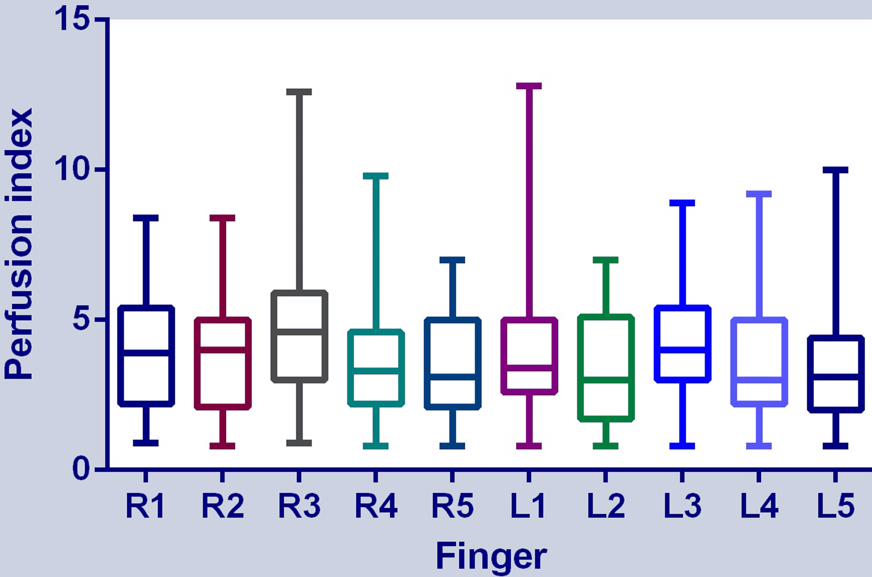
Cureus A Cross Sectional Study On The Agreement Of Perfusion Indexes Measured On Different Fingers By A Portable Pulse Oximeter In Healthy Adults
Measurement of the quality of tissue reperfusion by NIRS on the thenar eminence and the digital perfusion index.
. CEUS-derived parameters were reduced in COVID-19 associated AKI compared with healthy controls perfusion index 3415 vs. Stress perfusion and scar in recovered COVID-19 patients. Ventilation imaging using xenon-133 gas was performed per Nuclear Medicine physician request to confirm mismatched segmental perfusion defects in high probability results only in 4 patients after negative testing for COVID19.
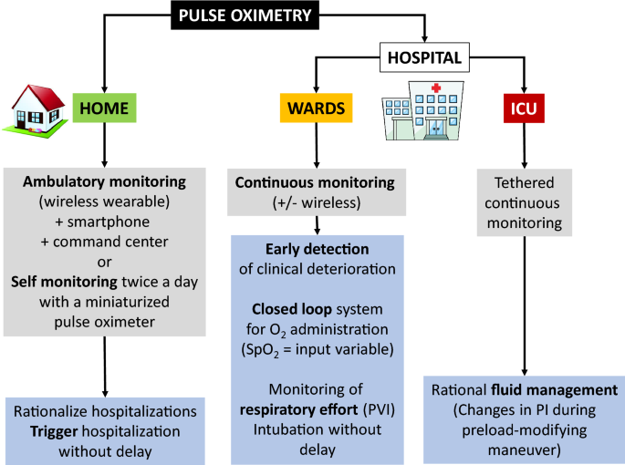
Microcirculation reactivity is measured for each patient with a laser speckle contrast imaging. This document supports the remote monitoring using pulse oximetry of patients with confirmed or possible COVID-19. The other 2 high probability results were confirmed with the clinical presentation and additional radiologic imaging.
Only wedge-shaped defects corresponded with PT two of 27 74. This may suggest that the shunt associated to the gasless lung parenchyma is not sufficient to exp. PBVPAenh 182 42 was lower than in healthy volunteers 27 139 P 002.
They found 27 54 of 50 patients to have a mix of restrictive and low diffusion patterns. Sublingual microcirculation was characterized by decreases in the proportion of perfused vessel and flow velocity along with high vascular densities. This last finding might be related to enhanced angiogenesis or hypoxia-induced capillary recruit.
The spectrum of perfusion abnormalities. Mean extent of perfusion defects was 361 172. COVID-19 patients showed an altered tissue perfusion.
Report of 3 autopsies from Houston Texas and review of autopsy findings from other United States cities. On March 19 2020 SNMMI released a statement responding to concerns regarding ventilationperfusion VQ lung scans and specifically the inherent risk of spread of COVID-19 to patients and staff related to the ventilation portion of this study. COVID-19 infection may lead to acute respiratory distress syndrome CARDS where severe gas exchange derangements may be associated at least in the early stages only with minor pulmonary infiltrates.
Perfusion index is an indication of the pulse strength at the sensor site. Perfusion abnormalities on dual-energy CT. Pertinently they reported an isolated decreased diffusing capacity in 13 26 of 50 patients raising the possibility that in addition to alveolar cell damage-related potential for fibrosis pulmonary vascular insult might also play a part.
Prone positioning may recruit gas exchange-efficient regions for typical acute respiratory distress syndrome ARDS and improve oxygenation. The emerging spectrum of cardiopulmonary pathology of the coronavirus disease 2019 COVID19. Preload modifying maneuvers such as tidal volume challenge can also be used in COVID-19 patients especially if the patient was in the gray zone of other dynamic tests.
In COVID-19 disease the infection of endothelial might cause an acute endothelial dysfunction. If a cardiac output monitor was not available the response to the passive leg raising test could be traced by measurement of the pulse pressure or the perfusion index. It is also a mainstay of treatment in COVID-19-related ARDS C-ARDS and reduces the need for intubation without any signal of harm In the early phase of COVID-19 hypoxemia may be caused by impaired regulation of.
COVID-19 is often associated with coagulopathy 14 15 that can lead to microemboli which in turn could redirect perfusion to lung regions. Perfusion abnormalities are common features of COVID-19 pneumonia including mosaic perfusion focal hyperemia in a subset of pulmonary opacities focal oligemia associated with a subset of peripheral opacities and rim of increased perfusion around an area of low perfusion hyperemic halo sign. PMC free article Google Scholar.
The present study retrospectively evaluated the prevalence and distribution of lung perfusion defects in early post-COVID-19 patients with hypoxia and was aimed to identify the risk factors for mismatched perfusion defects. 50 Best Overall Innovo iP900AP Deluxe Pulse Oximeter with Plethysmograph and Perfusion Index Buy Now On Amazon Price 3699 FDA-approved or for medical use Yes Why We Picked It Noteworthy. From left to right we show the first-pass perfusion images quantitative stress perfusion maps and free breathing-phase sensitive inversion recovery and motion corrected late gadolinium enhancement images PSIR MOCO LGE.
Dual-energy CT DECT can depict pulmonary perfusion by regional assessment of iodine uptake. Studies have shown that some patients with coronavirus disease 2019 COVID-19 and acute hypoxaemic respiratory failure have preserved lung compliance suggesting that processes other than alveolar damage might be involved in hypoxaemia related to COVID-19 pneumonia. Acute PT was present in 11 of 27 407 patients.
We retrospectively analyzed the. Perfusion-only scans were performed in consecutive patients from March 1 - August 31 2020 using 2-4 mCi for body mass index below or above 35 respectively of 99mTc-labeled macroaggregated albumin particles administered intravenously over 3. Perfusion Index or PI is the ratio of the pulsatile blood flow to the non-pulsatile static blood flow in a patients peripheral tissue such as in a fingertip toe or ear lobe.
Accordingly we examined a cohort of mechanically ventilated patients with severe COVID-19 pneumonia abnormalities focusing on 1 physiologic data 2 findings on computed tomographic pulmonary angiography CTPA 3 lung perfusion as demonstrated by dual-energy computed tomography DECT pulmonary blood volume maps and 4 hematologic tests evidence of. The purpose of this study was the analysis of pulmonary perfusion using dual-energy CT in a cohort of 27 consecutive patients. The purpose of this study was to define lung perfusion changes in COVID-19 cases with DECT as well as to reveal any possible links between perfusion changes and laboratory findings.
It is designed for patients in primary and community health settings and can also be used for patients who are at an early stage of the disease and sent home from AE or discharged following short hospital admissions. At that time many institutions opted not to perform ventilation studies. The PIs values range from 002 for very weak pulse to 20 for extremely strong pulse.
As we all do our best to avoid the spread of COVID-19 while continuing to provide the best care for our patients we would like to respond to concerns regarding ventilationperfusion VQ lung scans and the risk inherent in the VQ scan for spread of COVID-19 to patients and staff alike. Gas exchange in COVID-19 pneumonia is impaired and vessel obstruction has been suspected to cause ventilation-perfusion mismatch. Mean CT obstruction index was 26 54 out of 40.

Experience With A Perfusion Only Screening Protocol For Evaluation Of Pulmonary Embolism During The Covid 19 Pandemic Surge Journal Of Nuclear Medicine

Changes In Perfusion Index Induced By The Lung Recruitment Manoeuvre Download Scientific Diagram
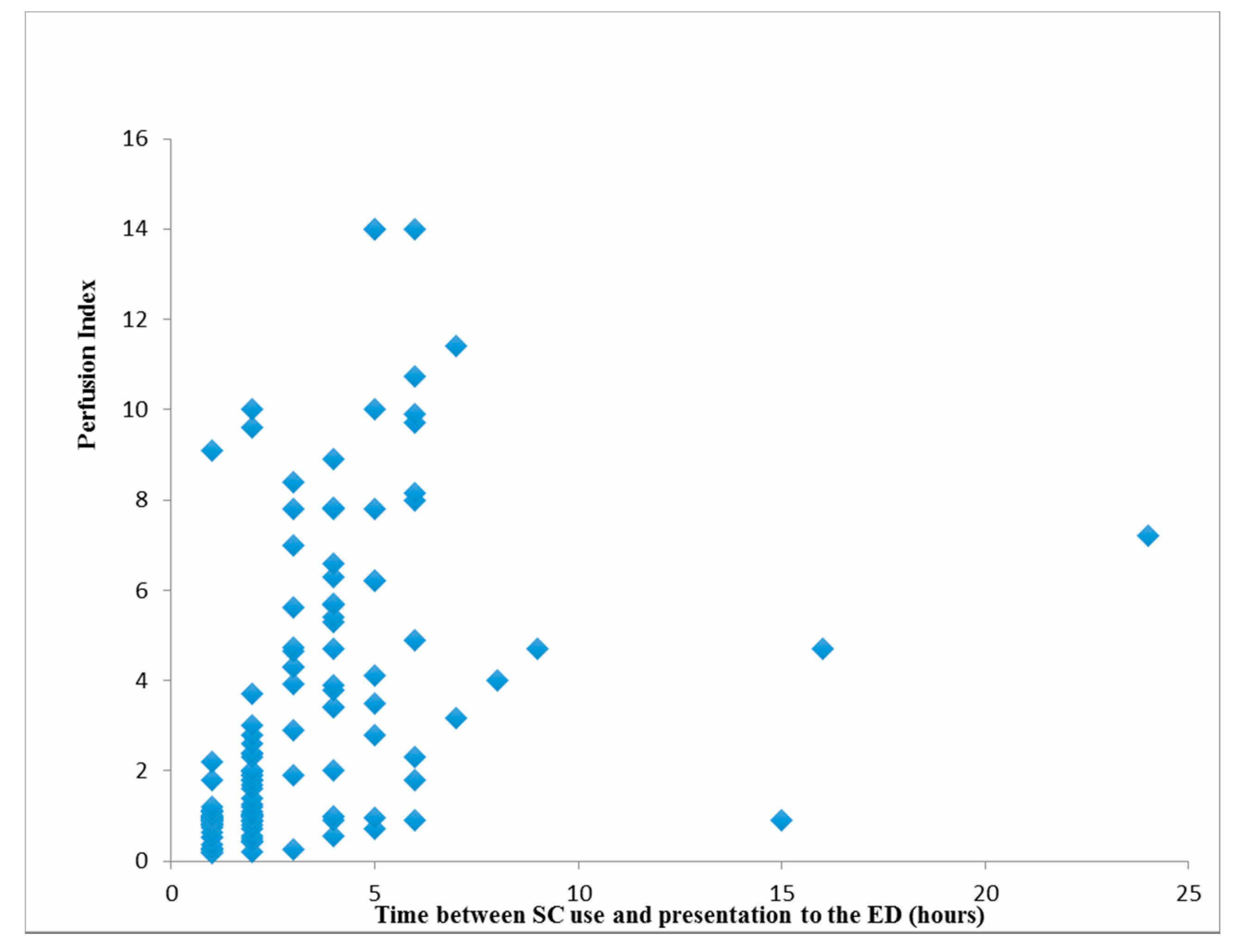
Medicina Free Full Text Perfusion Index Analysis In Patients Presenting To The Emergency Department Due To Synthetic Cannabinoid Use Html
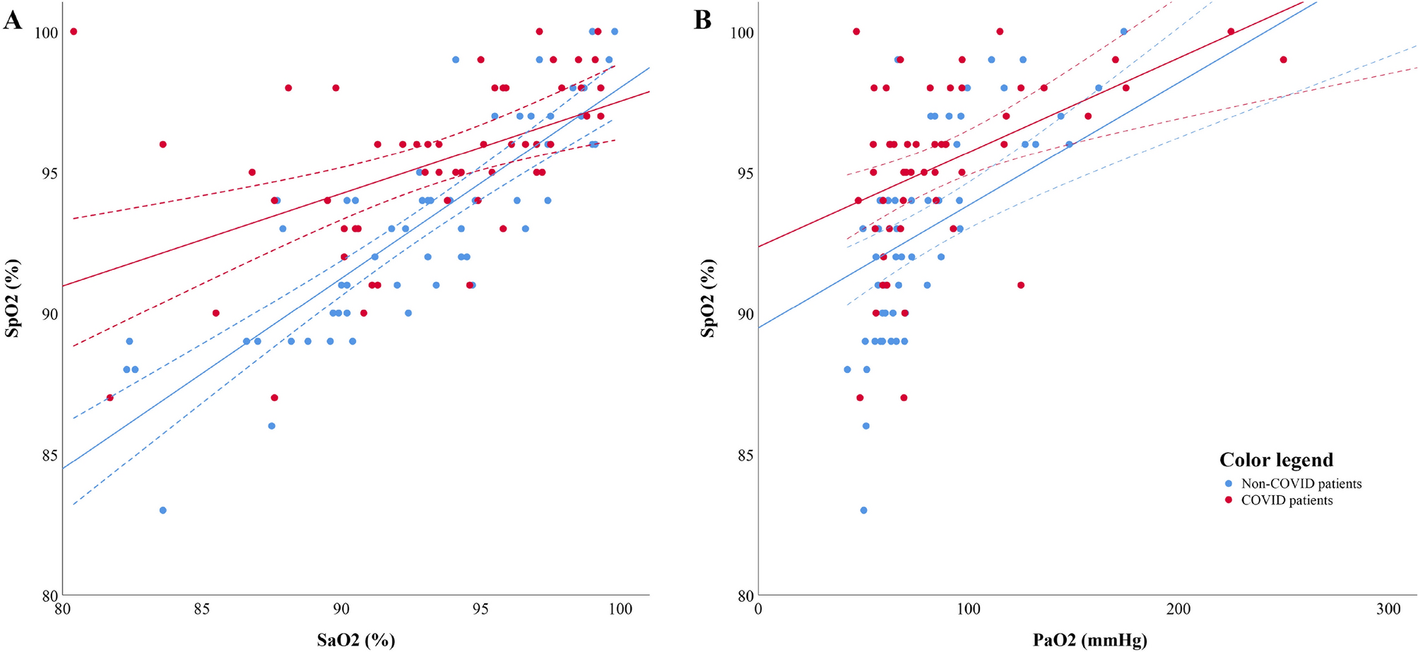
Impact Of Covid 19 On The Association Between Pulse Oximetry And Arterial Oxygenation In Patients With Acute Respiratory Distress Syndrome Scientific Reports

Covid 19 Pulse Oximeters In The Spotlight Springerlink
Invasive And Non Invasive Ventilation In Patients With Covid 19 03 08 2020

Changes In Perfusion Index Induced By The Lung Recruitment Manoeuvre Download Scientific Diagram

Impact Of Covid 19 On The Association Between Pulse Oximetry And Arterial Oxygenation In Patients With Acute Respiratory Distress Syndrome Scientific Reports
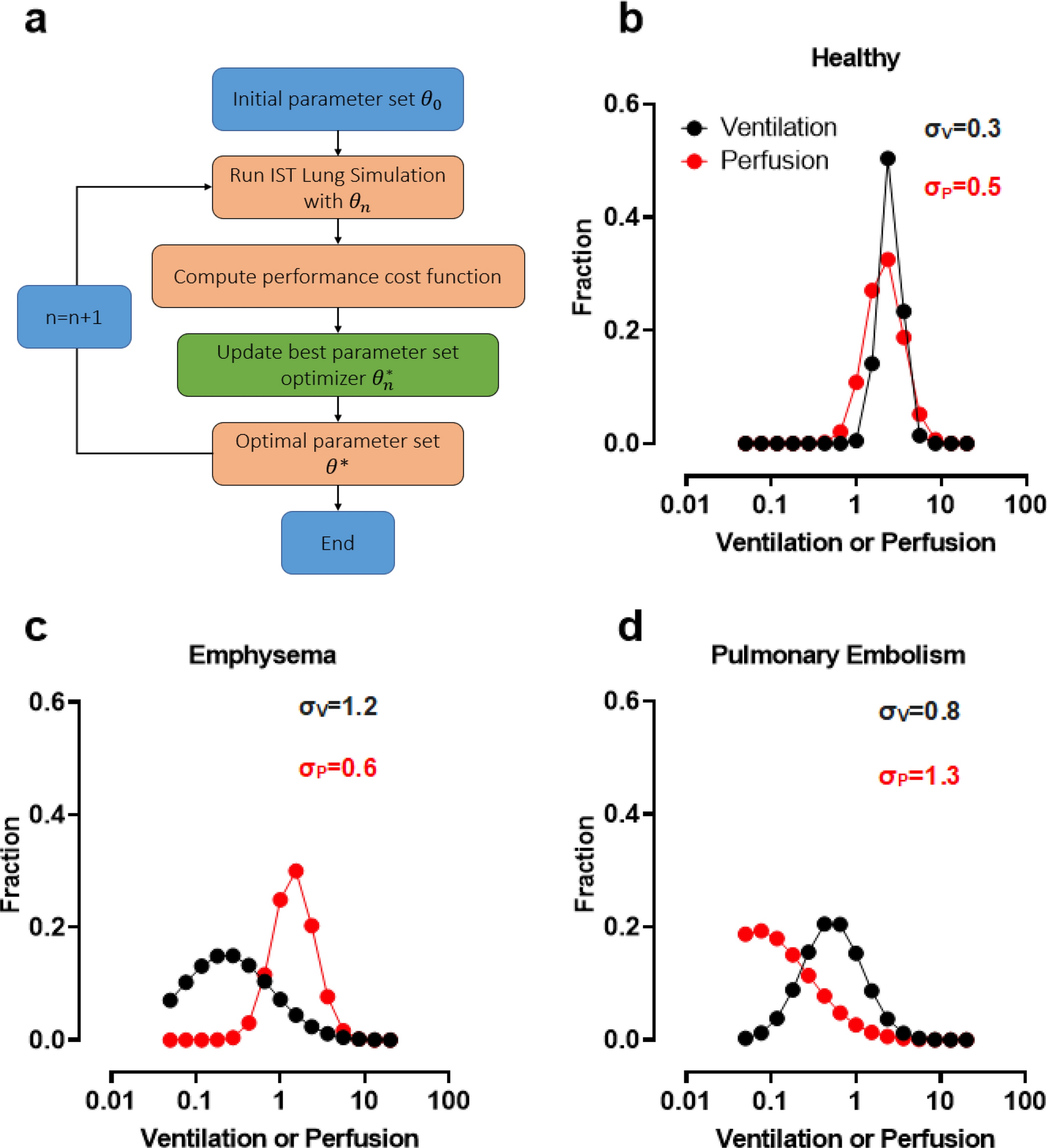
Simulation Based Optimisation To Quantify Heterogeneity Of Specific Ventilation And Perfusion In The Lung By The Inspired Sinewave Test Scientific Reports
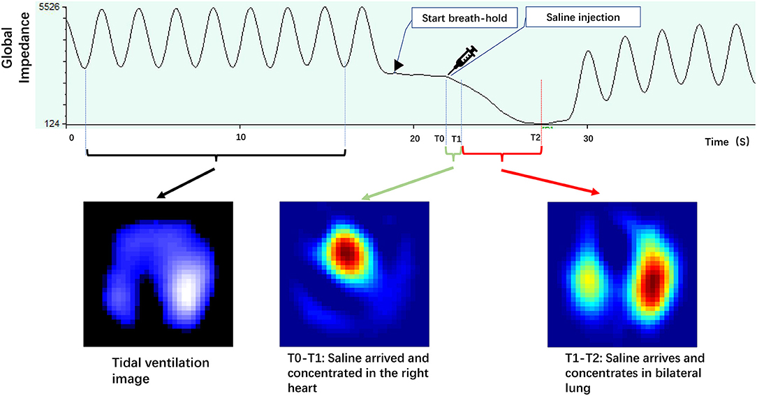
Frontiers Lung Perfusion Assessment By Bedside Electrical Impedance Tomography In Critically Ill Patients Physiology
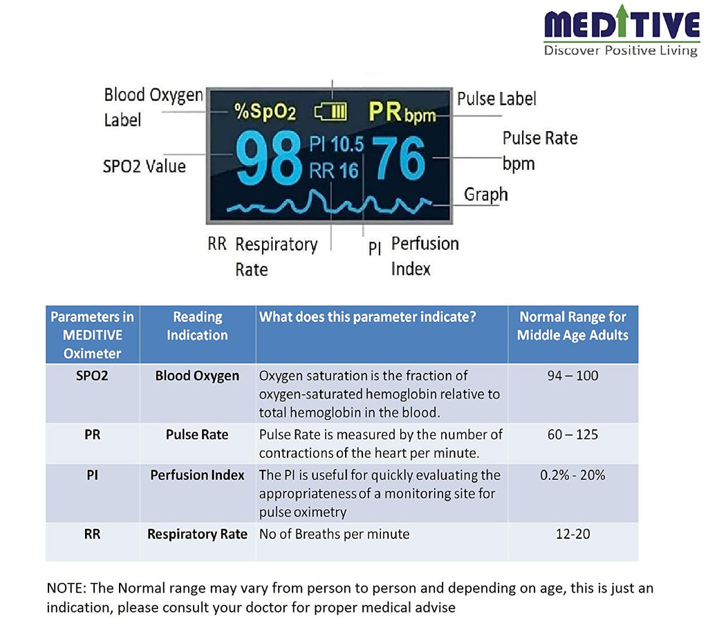
Pulse Oximeter Normal Clearance 53 Off Discoverlifeatl Com
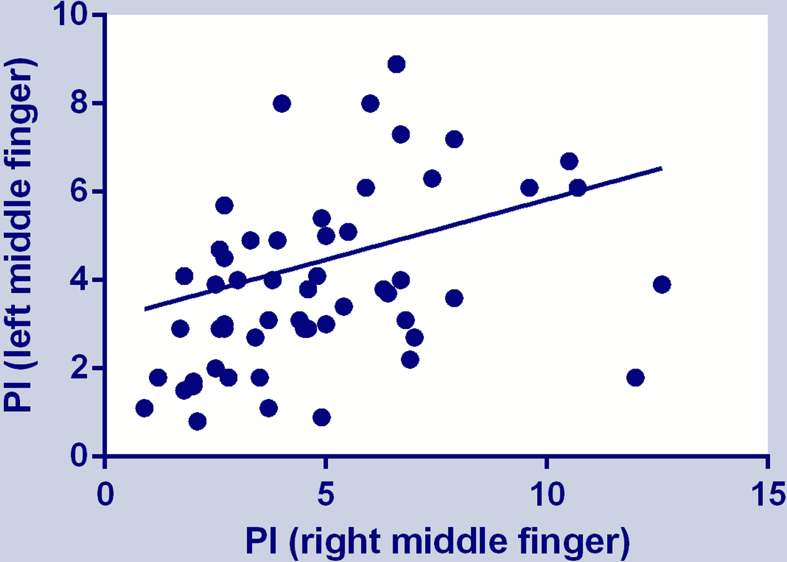
Cureus A Cross Sectional Study On The Agreement Of Perfusion Indexes Measured On Different Fingers By A Portable Pulse Oximeter In Healthy Adults

Ppg In Clinical Monitoring Sciencedirect

Relationship Between Changes In Perfusion Index And Changes In Stroke Download Scientific Diagram
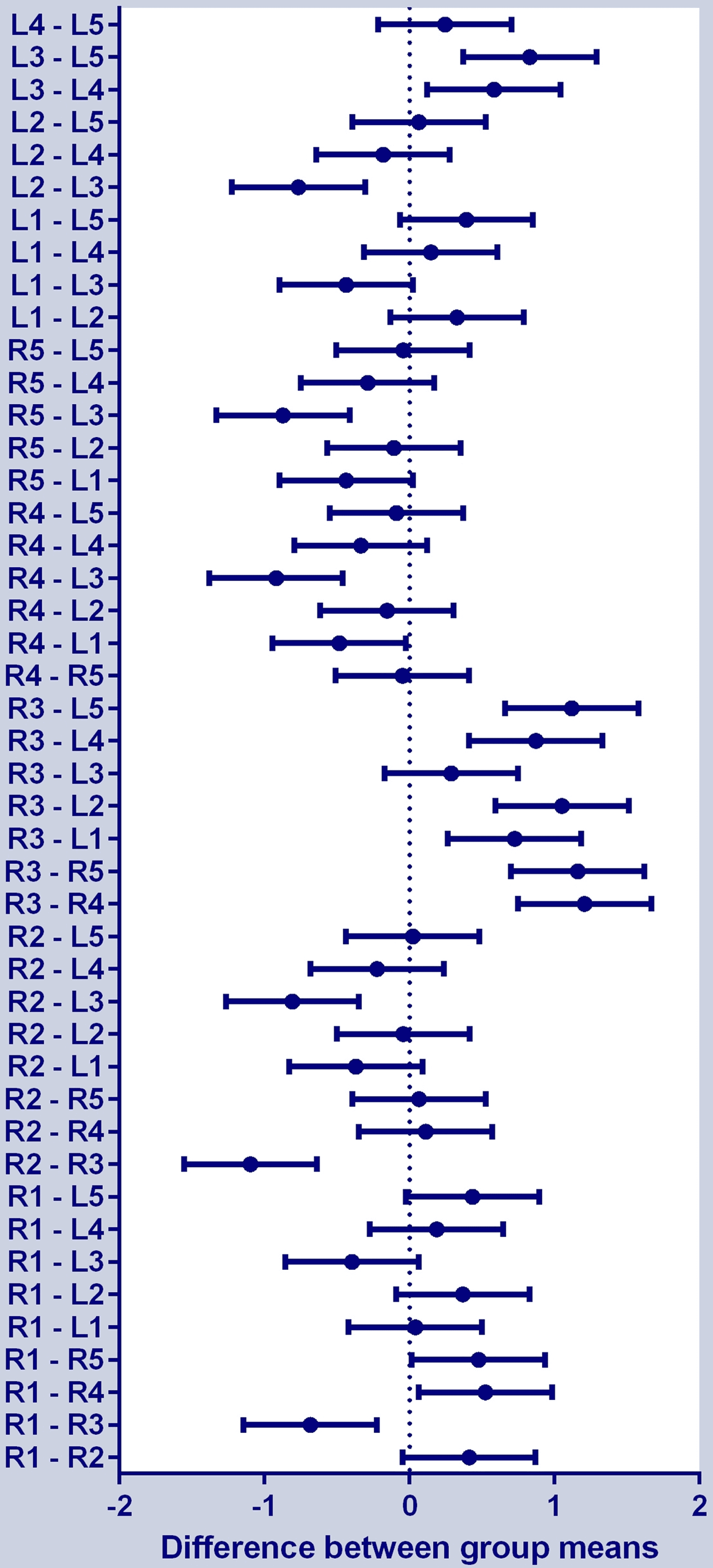
Cureus A Cross Sectional Study On The Agreement Of Perfusion Indexes Measured On Different Fingers By A Portable Pulse Oximeter In Healthy Adults

Perfusion Indices With And Without Nail Polish Download Table
Pedra Tissue Perfusion System Receives Fda Breakthrough Device Designation Daic
0 Response to "perfusion index covid"
Post a Comment